An aneurysm is the enlargement of an artery caused by weakness in the arterial wall. Often there are no symptoms, but a ruptured aneurysm can lead to fatal complications. An aneurysm refers to a weakening of an artery wall that creates a bulge, or distention, of the artery. Most aneurysms do not show symptoms and are not dangerous. However, at their most severe stage, some can rupture, leading to life-threatening internal bleeding.
.jpg)
A brain arteriovenous malformation (AVM) is a tangle of abnormal blood vessels connecting arteries and veins in the brain. The arteries are responsible for taking oxygen-rich blood from the heart to the brain. Veins carry the oxygen-depleted blood back to the lungs and heart. A brain AVM disrupts this vital process. An arteriovenous malformation can develop anywhere in your body but occurs most often in the brain or spine. Even so, brain AVMs are rare and affect less than 1 percent of the population. The cause of AVMs is not clear. Most people are born with them, but they can occasionally form later in life. They are rarely passed down among families genetically. Some people with brain AVMs experience signs and symptoms, such as headache or seizures. AVMs are commonly found after a brain scan for another health issue or after the blood vessels rupture and cause bleeding in the brain (hemorrhage). Once diagnosed, a brain AVM can often be treated successfully to prevent complications, such as brain damage or stroke.
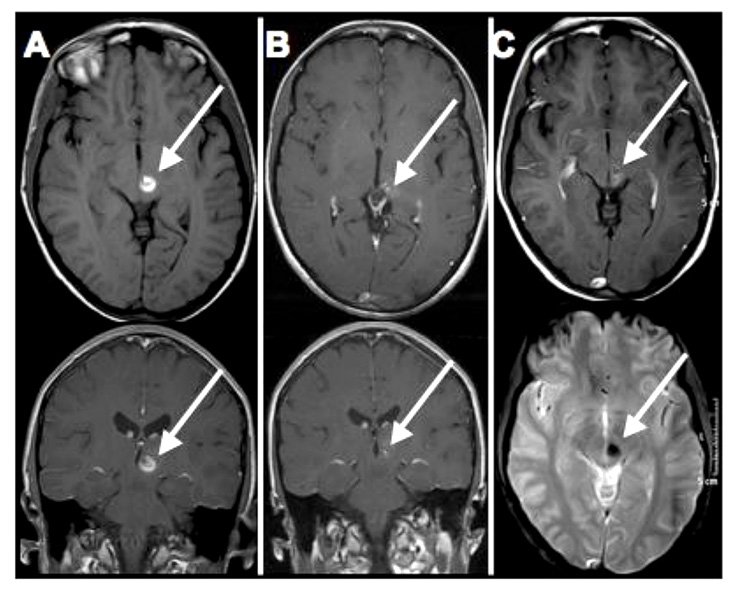
diagnose a cavernoma after you have had a seizure, a loss of function, or a surprise finding when we perform a magnetic resonance imaging scan for another reason. Cavernomas may have no symptoms but about one in three people with cavernous angiomas eventually develop symptoms, often between ages 20 and 40. The type, frequency and severity of symptoms often depend on the location of the cavernoma. Typical symptoms include:
.jpeg)
A carotid-cavernous sinus fistula (CCF) is an abnormal connection between an artery in your neck and the network of veins at the back of your eye. These veins at the back of your eye transport blood from your face and brain back to your heart and are located in small spaces behind your eyes called cavernous sinuses. Sometimes an abnormal channel forms between these veins and one of the internal or external carotid arteries that run up each side of your neck. This formation happens as a result of a small tear that sometimes occurs in one of the carotid arteries. If the tear occurs near the veins in the cavernous sinus, an abnormal channel may form between the artery and the network of veins, through which blood may flow. This is called a fistula. A fistula can raise the pressure in your cavernous sinuses, which may compress the cranial nerves located around the cavernous sinuses. This compression may damage the nerve function, which is to control your eye movements. These cranial nerves also allow you to experience sensation in parts of your face and head. The increased pressure caused by the fistula can also affect the veins that drain your eye. This can cause symptoms such as eye swelling and abnormal vision.
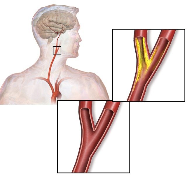
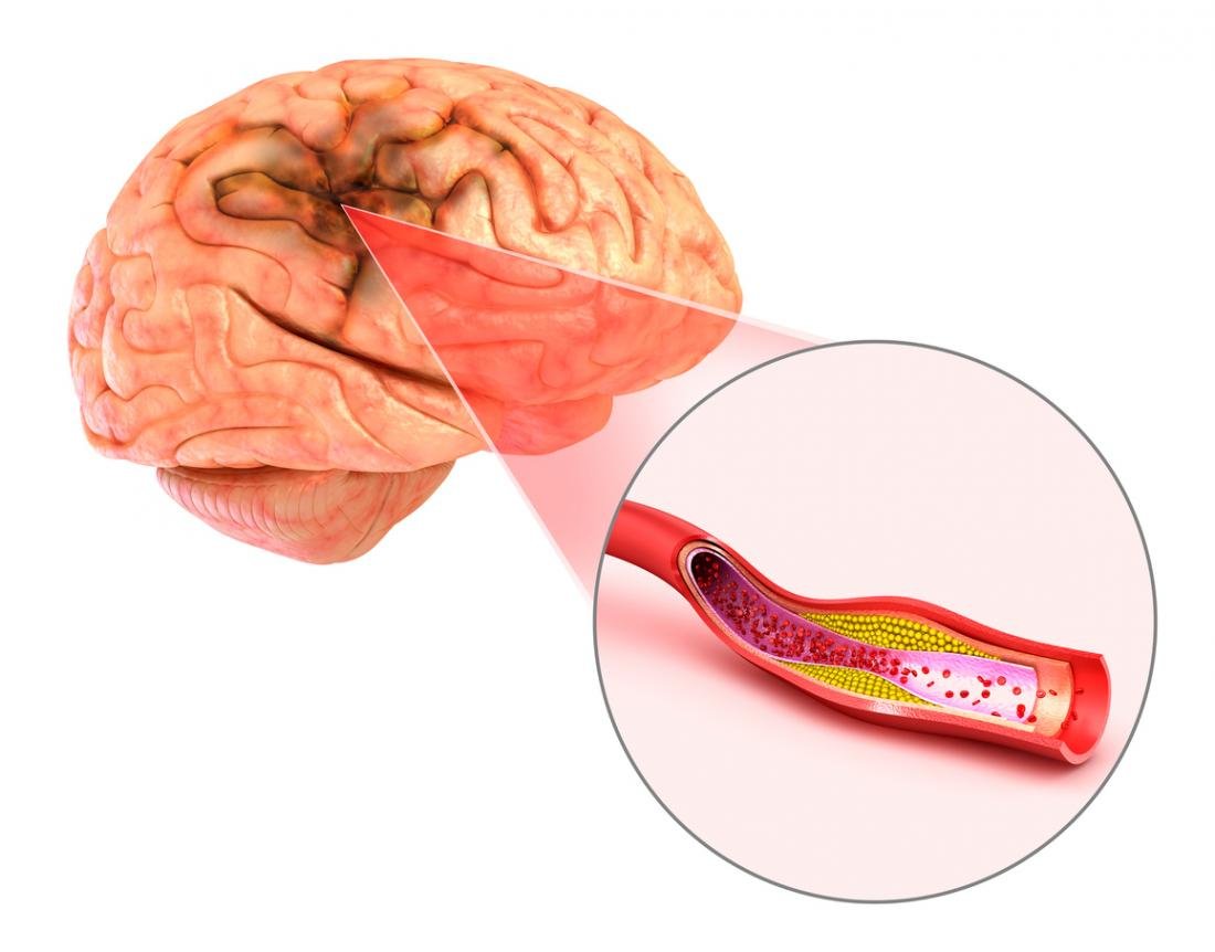
Carotid stenosis is a narrowing of the carotid arteries, the two major arteries that carry oxygen-rich blood from the heart to the brain. Also called carotid artery disease, carotid stenosis is caused by a buildup of plaque (atherosclerosis) inside the artery wall that reduces blood flow to the brain. Treatment aims to reduce the risk of stroke by controlling or removing plaque buildup and preventing blood clots. Blood supply of the brain To understand carotid stenosis, it is helpful to understand the circulatory system of the head and neck (see Anatomy of the Brain). The carotid artery begins at the aorta in the chest as the common carotid and courses up through the neck to the head. Place your hands on either side of your neck, and you can feel your pulse in your carotid arteries. Near the larynx, the common carotid divides into the external and internal carotid arteries. The external carotid arteries supply blood to the face and scalp. The internal carotid arteries supply blood to the brain. The most common location of atherosclerotic plaque buildup is the carotid bifurcation, where the common carotid divides into the internal and external carotid arteries.

Moyamoya disease is a rare blood vessel (vascular) disorder in which the carotid artery in the skull becomes blocked or narrowed, reducing blood flow to your brain. Tiny blood vessels then develop at the base of the brain in an attempt to supply the brain with blood. The condition may cause a ministroke (transient ischemic attack), stroke or bleeding in the brain. It can also affect how well your brain functions and cause cognitive and developmental delays or disability. Moyamoya disease most commonly affects children, but adults may have the condition. Moyamoya disease is found all over the world, but it's more common in East Asian countries, especially Korea, Japan and China. This may possibly be due to certain genetic factors in those populations.
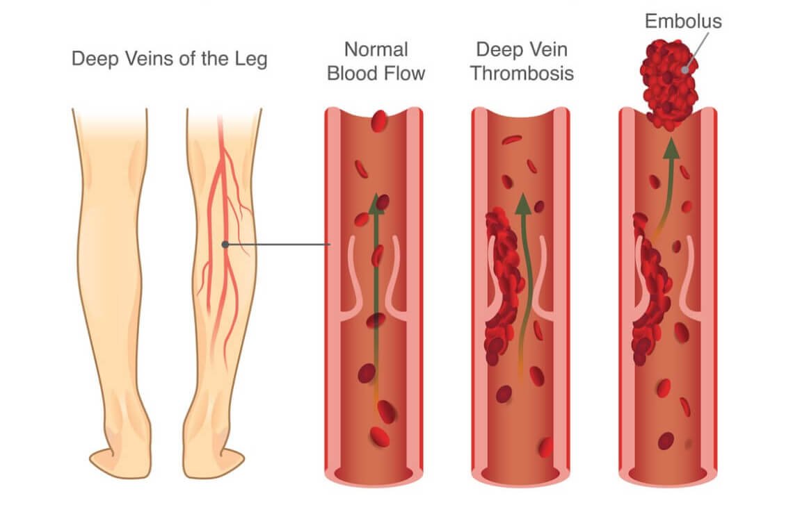
Venous thrombosis is thrombosis in a vein, caused by a thrombus (blood clot). A common form of venous thrombosis is deep vein thrombosis (DVT), when a blood clot forms in the deep veins. If the thrombus breaks off (it embolizes) and flows towards the lungs. It can become a pulmonary embolism (PE), a blood clot in the lungs. The conditions of DVT only, DVT with PE, and PE only are captured by the term venous thromboembolism (VTE). The initial treatment for venous thromboembolism is typically with either low-molecular-weight heparin (LMWH) or unfractionated heparin, or increasingly with directly acting oral anticoagulants (DOAC). Those initially treated with heparins can be switched to other anticoagulants (warfarin, DOACs), although pregnant women and some people with cancer receive ongoing heparin treatment. Superficial venous thrombosis only requires anticoagulation in specific situations, and may be treated with anti-inflammatory pain relief only.